Neuromuscular Disorders
IVIG Therapy for Neuromuscular Disorders
Specific neuromuscular disorders such as chronic inflammatory demyelinating polyneuropathy (CIDP) and Guillain-Barré Syndrome (GBS) can be treated with steroids, plasmapheresis (PP) and immunosuppressive drugs.  Many patients initially respond to these treatments, but develop resistance to the therapy or experience side effects causing the treatments to be stopped.
Researchers believe that intravenous immunoglobulin (IVIG) is longer lasting and provides patients with CIDP and GBS with a safer, more effective alternative to standard therapies. Â IVIG is a drug that has been used successfully to treat other immune-related diseases of the nervous system.

IVIG and plasmapheresis seem to be equally effective. IVIG is generally preferred to plasmapheresis because it is safer, more accessible and less invasive. Long term treatment with steroids can have serious side-effects. IVIG is effective in 70% to 90% of cases; however, most patients with initial improvement need long-term periodic doses of IVIG to maintain clinical stability.
If you have been diagnosed with CIDP or GBS and your physician recommends IVIG therapy, you will receive IVIG therapy on a regular basis. The infusion is usually given intravenously, which means through a needle directly into a vein at a doctor’s office, hospital, or infusion center. You may also be able to arrange to have your infusion at home.
Tolerability of IVIG is usually very good and adverse reactions are usually minor. The most common side effects are headache, nausea, chills, flushing, myalgia, hypotension, hypertension, chest discomfort, and fatigue. Infrequent adverse reactions include thromboembolic events, skin reactions, aseptic meningitis, renal tubular necrosis, and severe anaphylactic reaction.
Chronic Inflammatory Demyelinating Polyneuropathy (CIDP)
CIDP is an autoimmune disorder affecting the peripheral nerves. IG therapy helps reduce inflammation and slow disease progression, improving mobility and quality of life.
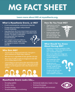
Myasthenia Gravis (MG)
In this condition, the immune system disrupts communication between nerves and muscles, causing weakness. IG therapy is used during severe exacerbations or to prepare for surgery, restoring strength and function.
Multifocal Motor Neuropathy (MMN)
MMN is a rare, progressive condition causing asymmetrical muscle weakness. IG infusion therapy can improve strength and delay further nerve damage.
Guillain-Barré Syndrome (GBS)
GBS occurs when the immune system attacks the peripheral nervous system, leading to paralysis. Early administration of IG therapy can speed recovery and reduce complications.
Please see our list of Neuromuscular Disease FAQ’s below for additional information:
Chronic Inflammatory Demyelinating Polyneuropathy (CIDP)
CIDP is a neurological disorder characterized by gradually (over a time period of months or years) increasing weakness of the legs and arms. It is caused by damage to the protective covering of the nerves, called myelin. Symptoms are variable and may be mild to debilitating. CIDP is treatable with IVIG home infusion therapy.
A patient with CIDP typically presents with difficulty walking which progressively worsens over months to years. Weakness, tingling or other abnormal sensations may also be experienced, and usually begin in the fingers or toes (on both sides of the body). Physical examination will usually show loss of reflexes, such as the knee and ankle jerk. Evaluation by a neurologist will often include an electrical test, a nerve conduction velocity-electromyography study. Your doctor may obtain blood and urine tests, including analysis of proteins, to look for causes of CIDP. CIDP is usually a chronic condition, which means that it may require long-term treatment.
Although the exact cause is unknown, it is believed that the immune system, which is normally protective, perceives myelin as foreign and attacks it. Just what starts this process is not clear. Some patients are found to have abnormal proteins in their blood, and these may facilitate damage. Over time, the destruction of myelin leads to weakness, numbness and tingling in the arms and legs.
Several treatment options are available. These include steroids, plasmapheresis and intravenous immune globulin (IVIG). The goals of treatment are to stop further damage to the myelin, prevent damage to the nerve fibers (axons), alleviate symptoms and prevent relapse, and if possible, create an environment that allows the myelin to regenerate. In patients with CIDP, IVIG has been shown to reduce disability, prevent relapse and even improve quality of life.[1]
References:
1. Donofrio PD, Bril V, Dalakas MC, et al. Safety and tolerability of immune globulin intravenous in chronic inflammatory demyelinating polyradiculoneuropathy. Arch Neurol. 2010 Sep;67(9):1082-8.
CIDP can be treated with steroids, plasmapheresis (PP) and immunosuppressive drugs. Many patients initially respond to these treatments, but develop resistance to the therapy or experience side effects causing the treatments to be stopped.
Researchers believe that intravenous immunoglobulin (IVIG) is longer lasting and provides patients with CIDP a safer, more effective alternative to standard therapies for the disease. IVIG is a drug that has been used successfully to treat other immune-related diseases of the nervous system.
IVIG and PP seem to be equally effective. IVIG is generally preferred to PP because it is safer, more accessible and less invasive. Long-term treatment with steroids can have serious side-effects. IVIG is effective in 70% to 90% of cases; however, most patients with initial improvement need long-term periodic doses of IVIG to maintain clinical stability[1].
At least five small randomized controlled studies have demonstrated the benefit of IVIG in the majority of patients with CIDP[2-7]. The ICE study is the largest and most recent trial of IVIG to treat CIDP[7]. The study not only confirmed the short-term efficacy of IVIG, but also demonstrated that a maintenance therapy can sustain improvement, increase quality of life over 12 months, and prevent further axonal degeneration[7-10]. The ICE study advocates IVIG as a first-line therapy, and has led to FDA approval of one brand of IVIG. The study has also shown that repeated therapy is usually needed to improve symptoms and then to maintain that improvement. Typical doses recommended repeat every 1 to 6 weeks. Interestingly, a large part of the IVIG treatment responsive patients were able to wean off of therapy after 24 weeks without showing a relapse before the study period ended. So, the amount of medication and the frequency of the need for repeated doses can vary widely from patient to patient and is usually determined by the patient’s response to the drug and their physician’s experience with other similar patients in the past.
References:
1. Van Doorn PA. Treatment of patients with chronic inflammatory demyelinating polyneuropathy. Rev Neurol (Paris). 1996;152:383-386. 2.Mendell, J. R. et al. Randomized controlled trial of IVIg in untreated chronic inflammatory demyelinating polyradiculoneuropathy. Neurology 56, 445-449 (2001). 3.Hahn, A. F. Treatment of chronic inflammatory demyelinating polyneuropathy with intravenous immunoglobulin. Neurology 51 (6 Suppl. 5), S16-S21 (1998). 4.Vermeulen M. et al. Intravenous immunoglobulin treatment in patients with chronic inflammatory demyelinating polyneuropathy: a double blind, placebo controlled study. J. Neurol. Neurosurg. Psychiatry 56, 36-39 (1993). 5.Dyck, P. J. et al. A plasma exchange versus immune globulin infusion trial in chronic inflamatory demyelinating polyradiculoneuropathy. Ann. Neurol. 36, 838-845 (1994). 6.van Doorn, P A., Brand, A., Strengers, P F., Meulstee, J. Vermeulen, M. High-dose intravenous immunoglobulin treatment in chronic inflammatory demyelinating polyneuropathy: a double-blind, placebo-controlled, crossover study. Neurology 40, 209-212 (1990). 7.Hughes, R. A. C. et al. Intravenous immune globulin (10% caprylate-chromatography purified) for the treatment of chronic inflammatory demyelinating polyradiculoneuropathy (ICE study): a randomised placebo-controlled trial. Lancet Neurol. 7, 136-144 (2008). 8.Merkies, I. S. et al. Health-related quality-of-life improvements in CIDP with immune globulin IV 10%: the ICE Study. Neurology 72, 1337-1344 (2009). 9.Bril, V. et al. Electrophysiologic correlations with clinical outcomes in CIDP. Muscle Nerve 42, 492-497 (2010). 10.Latov, N. et al. Timing and course of clinical response to intravenous immunoglobulin in chronic inflammatory demyelinating polyradiculoneuropathy. Arch. Neurol. 67, 802-807 (2010).
IVIG is usually given initially at a dose of 0.4 g/kg per day for 5 days, followed by 1.0 g/kg or less as a single infusion in monthly or bimonthly intervals. The response is assessed after 1 to 2 months[11]. A weekly dosing schedule may also be useful for maintenance therapy in select cases[12].
References:
11. Hahn AF, Bolton CF, Zochodne D, Feasby TE. Intravenous immunoglobulin treatment in chronic inflammatory demyelinating polyneuropathy: a double-blind, placebo-controlled, cross-over study. Brain. 1996;119(pt 4):1067-1077 12. Dyck PJ, Litchy WJ, Kratz KM, et al. A plasma exchange versus immune globulin infusion trial in chronic inflammatory demyelinating polyradiculoneuropathy. Ann Neurol. 1994;36:838-845.
If you have been diagnosed with CIDP and your physician recommends IVIG therapy, you will receive IVIG therapy on a regular basis. The infusion is usually given intravenously, which means through a needle directly into a vein at a doctor’s office, hospital, or infusion center. You may also be able to arrange to have your infusion at home.
Tolerability of IVIG is usually very good and adverse reactions are usually minor. The most common side effects are headache, nausea, chills, flushing, myalgia, hypotension, hypertension, chest discomfort, and fatigue. Infrequent adverse reactions include thromboembolic events, skin reactions, aseptic meningitis, renal tubular necrosis, and severe anaphylactic reaction.
Dermatomyositis (DM)
Dermatomyositis is an uncommon muscle disease that is accompanied by a skin rash. It affects adults and children alike. In adults, dermatomyositis usually occurs from the late 40s to early 60s; in children, the disease most often appears between 5 and 15 years of age. Dermatomyositis affects more females than males. DM is treatable with IVIG home infusion therapy.
• Difficulty swallowing or talking
• Shortness of breath
• Weight loss
• Low-grade fever
• Sensitivity to light
• Calcium deposits under the skin
• Nail changes
AlopeciaÂ
The exact cause of dermatomyositis is unknown, but the disease is similar to other autoimmune disorders, in which your immune system attacks itself. Research is taking place to identify other factors that may play a part in its development, some of which are an infection, underlying skin cancer(more likely in elderly) and genetic predisposition.
There is no cure for dermatomyositis. The primary aim of treatment is to control the skin condition and the muscle strength and function. Treatment options include antimalarial medications, corticosteroids to reduce inflammation, immunosuppressant medications, intravenous immunoglobulins(IVIG) to reduce the immune response, pain relieving medications and steroid-sparing agents. Other important measures in the management of dermatomyositis include physical therapy, speech therapy, and dietary counseling.
The mainstay of dermatomyositis treatment usually involves oral corticosteroids to slow down the rate of disease progression. Immunosuppressive medications may also be used in conjunction with corticosteroids. These medicines all have significant side effects and often a less than adequate response is achieved with this conventional therapy.
IVIG is an effective additional therapy for patients with dermatomyositis who fail to respond to conventional therapy or who experience unacceptable side effects. A summary of clinical trials shows an overall response rate of 80% at about 2 months, with maximal response at 4 months. Most patients require ongoing IVIG therapy in addition to conventional treatments given at lower and better-tolerated dosages.
IVIG is recommended to be given monthly at a dose of 1-2 g/kg administered over 5 days. Usually, the effects of IVIG can last up to a month after each administration. IVIG is given as an infusion into a vein over a period of time, usually from 4 to 6 hours.
Skin reactions to IVIG are uncommon. Of all the reported rashes, a blistering type of eczema is the most common. [1] It often begins at about 8 to 10 days after exposure to IVIG. The rash characteristically begins as small itchy blisters on the palms and/or soles that then extends to the rest of the body. The affected individual may become red all over and itchy. Switching the type of IVIG may eliminate the reaction.
The skin lesions often resolve within a period of 1 to 4 weeks. The use of steroids controls symptoms and may speed recovery.
Reference:
1. Vecchietti, G., et al., Severe eczematous skin reaction after high-dose intravenous immunoglobulin infusion: report of 4 cases and review of the literature. Arch Dermatol, 2006. 142(2): p. 213-7.
Guillian-Barre Syndrome (GBS)
GBS is a neurological disorder characterized by gradually increasing weakness of the legs and arms over a period of days to weeks. It is caused by damage to the protective covering of the nerves, called myelin. Symptoms are variable and may be mild to debilitating. GBS is treatable with IVIG home infusion therapy.
A patient with GBS typically presents with muscle weakness and abnormal sensations in the feet and legs which may ascent to the arms and upper body in a rapid fashion. Physical examination will usually show loss of reflexes, such as the knee and ankle jerk. Unlike GBS, patients with CIDP typically experience symptoms over a period of several months to years, while symptoms in patients with GBS usually progress more quickly and people reach a critical period of illness within 2 to 3 weeks or less. In severe cases, breathing difficulty and/or paralysis may occur. For these patients hospitalization and placement on a ventilator may be required.
Although the exact cause is unknown, it is believed that the immune system, which is normally protective, perceives myelin as foreign and attacks it. Just what starts this process is not clear. Some patients are found to have abnormal proteins in their blood, and these may facilitate damage. The acute onset of symptoms helps to differentiate it from other diseases.
Plasmapheresis or IVIG is usually given as soon as a diagnosis of GBS is established in an effort to stop the ascending paralysis before it compromises the patient’s respiration. In some patients, only a few courses of IVIG are needed to control symptoms and avoid significant disability. Clinical evidence exists to support the use of IVIG to treat patients with GBS.[1]
Reference:
1. Donofrio PD, Berger A, Brannagan TH 3rd, et al.Consensus statement: the use of intravenous immunoglobulin in the treatment of neuromuscular conditions report of the AANEM ad hoc committee. Muscle Nerve. 2009 Nov;40(5):890-900.
The management of GBS consists of both supportive and immunomodulatory treatments, of which intravenous immunoglobulin (IVIG) and plasmapheresis (PP) are considered most effective. A number of randomized, controlled studies have shown IVIG to be at least as effective as plasmapheresis in the treatment of GBS, and in some cases, superior. The choice of treatment for individual patients is undertaken by the patient’s physician, based on the patient’s condition, and sometimes on the hospital at which the patient is being treated.
Not all hospitals have the equipment and personnel required to offer PP, besides which the method is difficult to administer to elderly patients and those with poor venous access. IVIG is more convenient to administer and receive, and has therefore often replaced plasmapheresis as the preferred treatment. It can be administered at most hospitals and can be administered as a 4 to 6 hour infusion. Also, IVIG does not remove other useful components that are in the blood and can be less costly and has less problems than does PP.
Moreover, IVIG has been found to be safer than PP, having a lower frequency of complications. IVIG has also been found to be effective and safe in the treatment of pediatric patients with GBS[1,2]. Thus, its efficacy, safety, and availability make it the treatment of choice in many patients with GBS.
Some evidence suggests that in select patients who do not respond initially to IVIG, a second dose may be beneficial.[3]Â However, this is not currently standard therapy and warrants further investigation.
References:
1.Hughes RA, Wijdicks EF, Barohn R, et al. Practice parameter: immunotherapy for Guillain-Barré syndrome: report of the Quality Standards Subcommittee of the American Academy of Neurology. Neurology. Sep 23 2003;61(6):736-40.
2.Yata J, Nihei K, Ohya T, et al. High-dose immunoglobulin therapy for Guillain-Barré syndrome in Japanese children. Pediatr Int. Oct 2003;45(5):543-9.
3. Van Doorn PA, Kuitwaard K, Walgaard C, et al. IVIG treatment and prognosis in Guillain-Barré syndrome. J Clin Immunol. May 2010;30 Suppl 1:S74-78.
demyelinating polyradiculoneuropathy. Arch. Neurol. 67, 802-807 (2010).
Usually, the starting dose is 2 grams per kilogram of patient weight, given in 5 divided dosages over 5 days. Further doses are based on patient response. The IVIG effect on GBS may be seen in a few days and may last for several weeks to months. Some patients may need further doses in the future if symptoms return.
IVIG is given as an intravenous drip.
In general, IVIG is considered safe. Patients may experience mild side-effects including headaches, stiffness of the neck, nausea, dizziness, vomiting, chills, fever, low blood-pressure and arrhythmia for up to 48 hours after being treated, or in the initial stages of the treatment. These symptoms disappear after a few hours or days. If they occur during the treatment, they can be minimized by reducing the rate of IVIG infusion.
The treatment can also trigger allergic reactions, such as a rash on the palms of the hands. Serious complications, seldom seen in otherwise healthy patients, include kidney damage and the formation of blood clots.
Multifocal Motor Neuropathy (MMN)
Multifocal motor neuropathy (MMN) is a rare condition in which multiple motor nerves are attacked by one’s immune system. This causes weakness without loss of sensation. The specific nature of the attack is unique and perplexing, since motor and sensory fibers are intermingled within the nerve trunks of the arms and legs, but only the motor nerves become involved.[1] MMN is treatable with IVIG home infusion therapy.
Reference:
1. Katz J, Lewis R. Multifocal Motor Neuropathy. Neuropathy Action Foundation, 2013.
By definition MMN causes weakness. There is essentially no numbness or tingling, and pain is not a significant factor. MMN usually develops asymmetrically and tends to begin in the hands. Frequently, the weakness can be recognized as fitting a specific nerve territory. For example, someone who develops a wrist and finger drop has a problem in the radial nerve, and when new weakness occurs, a different nerve is likely to be involved. The attack on multiple single nerves is called multiple mononeuropathy syndrome. This pattern can be seen in other diseases, but MMN is the only condition where the attack is isolated to the motor nerve fibers. It follows that this is the most important clinical finding to diagnose MMN.
Patients with MMN can have other symptoms including twitching, or small random dimpling of the muscle which neurologists call fasciculations. Fasciculations are the spontaneous firing of a motor unit (the collection of all the muscle fibers that are innervated by a single motor neuron). Fasciculations are also characteristic of ALS (amyotrophic lateral sclerosis) and this is one reason many patients with MMN are initially misdiagnosed with ALS. Other lower motor neuron signs such as atrophy, decreased tone and absent reflexes occur in both diseases, although in MMN these tend to affect the territories of single nerves, while in ALS they affect all of the muscles in the limb. In contrast to MMN, patients with ALS may have “upper motor neuron signs” (UMN) caused by damage to motor control in the brain. These signs include increased muscle tone and reflexes and pathologic reflexes such as a Babinski sign.[1]
Reference:
1. Katz J, Lewis R. Multifocal Motor Neuropathy. Neuropathy Action Foundation, 2013.
which means that it may require long-term treatment.
The diagnosis of MMN depends on demonstrating that a patient has a purely motor disorder affecting individual nerves, with criteria designed to differentiate the disorder from ALS (amyotrophic lateral sclerosis), the Lewis-Sumner syndrome variant of chronic inflammatory demyelinating polyneuropathy (CIDP) (similar to MMN but usually with significant sensory loss), and “vasculitis” (a type of multiple mononeuropathy syndrome caused by inflammatory damage to the blood vessels in nerves that also causes sensory and motor symptoms).[1]
Reference:
1. Katz J, Lewis R. Multifocal Motor Neuropathy. Neuropathy Action Foundation, 2013.
It is now established that intravenous immunoglobulin (IVIG) provides benefit to patients with MMN. IVIG can lead to improvement in most patients with MMN, with the response varying from minimal to very large. The treatment usually does not completely reverse all of the symptoms and those patients who do respond will require repeated treatments to keep their improvements. The exact timing and dosing need to be individualized and there is no single formula for success. Dosing may need to be adjusted if the response begins to wear off, or if symptoms worsen despite maintenance therapy.
IVIG is not a cure for MMN and currently no other therapy has proven effective. It is fairly clear that corticosteroids are ineffective and can actually make the disease worse. Other immunosuppressants have been used, but have greater side effects and risks. For example, there are a number of reports suggesting that cyclophosphamide controls the disease in some patients, while results for Rituximab are not encouraging. It is clear that newer therapies are needed, and many investigators around the world are working towards a better understanding of MMN and the development of more definitive treatments.[1]
Reference:
1. Katz J, Lewis R. Multifocal Motor Neuropathy. Neuropathy Action Foundation, 2013.
2010 Sep;67(9):1082-8.
Myasthenia Gravis (MG)
The term myasthenia gravis comes from the Greek and Latin words meaning “grave muscle weakness.” MG is believed to be a chronic autoimmune neuromuscular disorder characterized by weakness of the voluntary muscle groups, which are the muscles we use to perform physical activities. For reasons that are not clearly understood, the immune system of patients with MG makes antibodies against the receptor sites of the neuromuscular junction. Certain muscles such as those that control eye and eyelid movement, facial expression, chewing, talking and swallowing are often, but not always, affected by the disorder. The muscles that control breathing and neck and limb movements may also be involved. MG is treatable with IVIG home infusion therapy.
A patient with MG typically presents with muscle weakness. However, because this is a common symptom, diagnosis of MG may be delayed. A physician will perform a detailed history and physical examination, and look for specific muscle impairment (such as eye movements) as well as weakness that comes on with activity and improves following rest. Diagnosis can be confirmed with several tests including blood tests for acetylcholine antibody receptors and anti-MuSK antibodies, electromyography, nerve conduction studies and/or a tensilon test to measure effectiveness of the drug to alleviate fatigue for a short period of time.
MG is caused by a defect in the transmission of nerve impulses to muscles, but often the cause of contraction of MG for a particular patient is unknown. It occurs in all ethnic groups and both genders. It most commonly affects young adult women (under 40) and older men (over 60), but it can occur at any age. The thymus gland, part of the immune system, is abnormal in most MG patients. Some people with MG have benign tumors of the thymus gland called thymomas. Unfortunately, the relationship between the thymus gland and MG is not fully understood.
The goal of MG treatment is to remove/reduce antibodies and inhibit the binding of antibodies through various medications and treatments. There is no known cure for MG, but there are treatments that allow many people with MG to lead full lives. Common medications used to treat MG include pyridostigmine, prednisone, mycophenolate mofetil, azathioprine and cyclosporine. Thymectomy, or surgical removal of the thymus gland, may be an effective treatment for some patients. Clinical evidence exists to support the use of intravenous immunoglobulin (IVIG) infusions in patients with MG, especially during an exacerbation to prevent complications.
Please note that the content on this website is not intended to be a substitute for professional medical advice, diagnosis, or treatment. KabaFusion does not recommend or endorse any specific tests, products, procedures, or other information mentioned on this website.
Experience the difference with KabaFusion's Patient-Focused Care
Connect with a representative to learn more about infusion therapy treatment options or find a KabaFusion Pharmacy near you.
Call Now: 877.577.IVIG (4844)